医療機関のみなさまへ
現在70病院に導入をいただいております。
導入施設は、国立/都立/市立などの公立病院のほか、総合病院・クリニック・PET施設・IVR施設など多彩で、多様なニーズにお応えすることができます。
無痛MRI乳がん検診(ドゥイブス・サーチ)の導入をご検討の施設様は下記フォームからご連絡ください。
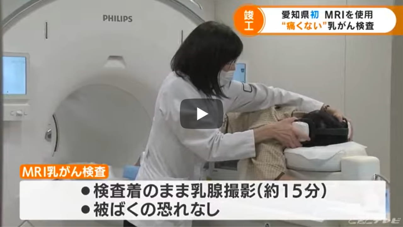
メディアからも注目されています。
医療機関から選ばれる
7つのポイント
無痛MRI乳がん検診
01
画質はピカイチ。
DWIBS法の開発者が
責任を持って設定します
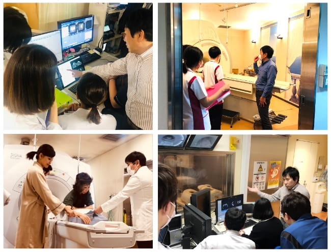
貴院に直接訪問して、テストスキャンを行い、画質を調整します。サービス開始後も、MRI装置への付着物やコイルの故障などにより画質は劣化しますので、診断時にわずかでも不良の兆候を見つけたら、本部から病院に連絡します。
これを継続的に繰り返すことにより、安心した画質を得ることができます。
DWIBS画質認証マーク
この認定マークを病院やウェブサイト、ポスター・DMなどに使用していただくことができます。
受診者さんが「病院を選ぶ」ときの一番の関心は、画質が良いのか、精度が高いのかということです。認定マークは、そのときの判断に役立てていただけます。
02
ウェブ予約システム自動付帯
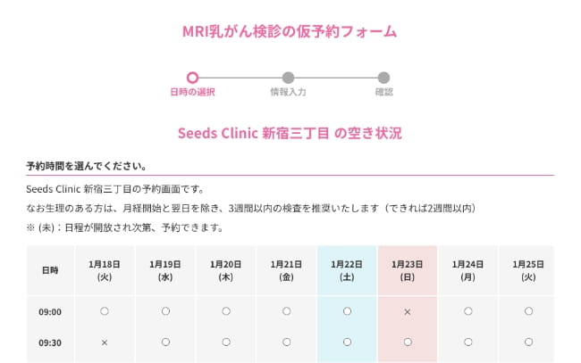
高いページビュー数と予約率
当社ホームページの月間ビュー数は平均月8万PV、10月のピンクリボン運動月間は20万PVほどになります。痛くない乳がん検診として認知もされており、信頼感により高い予約率を誇ります。
病院のウェブ予約システムとの併用も可能
病院独自のウェブ予約システムを持っていらっしゃるところは、独自のものだけをお使いいただくこともできますが、併用もできます。(上記理由により予約数が多くなるので、併用を強くおすすめしています)
マスメディア・SNSでの紹介
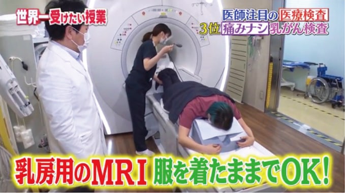
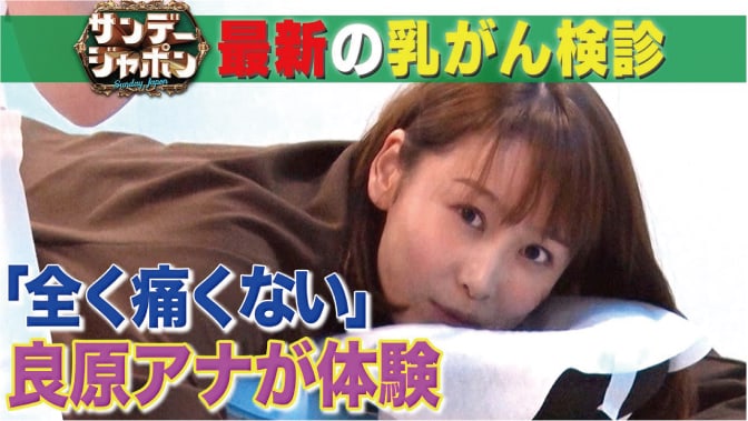
当社の取り組みは、記者・メディア関係の皆様から高い評価を得ており、このために、新聞、雑誌、またテレビでも扱っていただけます。例えばテレビではこれまでに、「NHKニュース」「サンデージャポン」「世界一受けたい授業」などの番組でご紹介をいただきました。
03
効果的なアンケートの
実施による高いリピート率
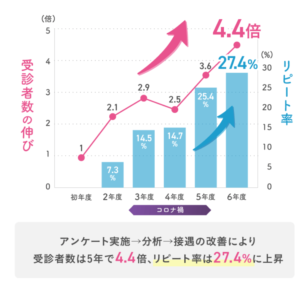
毎年リピート率が増える
このグラフは、提携病院のひとつの例です。導入から5年間で、受診者数4.4倍、リピート率は実に27.4%まで高まりました。続ければ続けるほど安定した受診者数が望めることがわかります。
接遇指導とアンケート
このためのポイントが接遇です。このサービスでは、まずテストスキャン時に、どのような接遇を行うと評判が良くなるのかについて、これまで得られたノウハウをもとに詳しく直接お教えします。
そのうえで、アンケート実施結果をお送りいただき、私共が分析してフィードバックすることで、顧客推奨度をどんどん高めています。このことにより、さらに推奨度が上がり、リピーターの獲得に繋がります。
04
非造影MRI検査の
スペシャリストによる読影
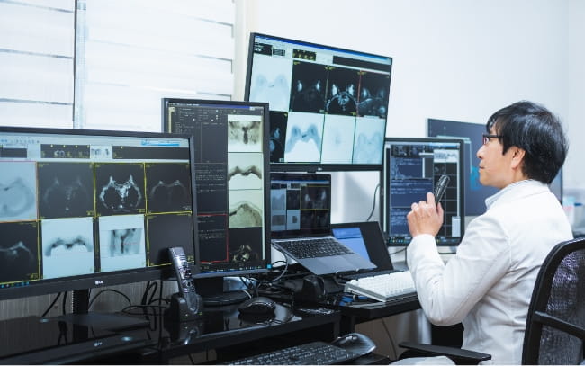
専門の読影医が診断
非造影MRIによる乳がん検診は新しいジャンル。特別な教育を受けた読影医が診断します。常勤の読影医は忙しく、検診まで継続的にサポートすることは大変難しいです。このサービスでは、完全に読影を請け負います。
レポートチェックと、統一感のある文体
また内部でのレポートチェックは3回行い、文字の誤りなどを可能な限り少なくしています。さらに、読影医によってまちまちになりがちな遠隔読影の欠点を排除し、文量や文体も統一感がでるように常に修正を行っています。
05
乳腺外科のない病院でも
導入可能です
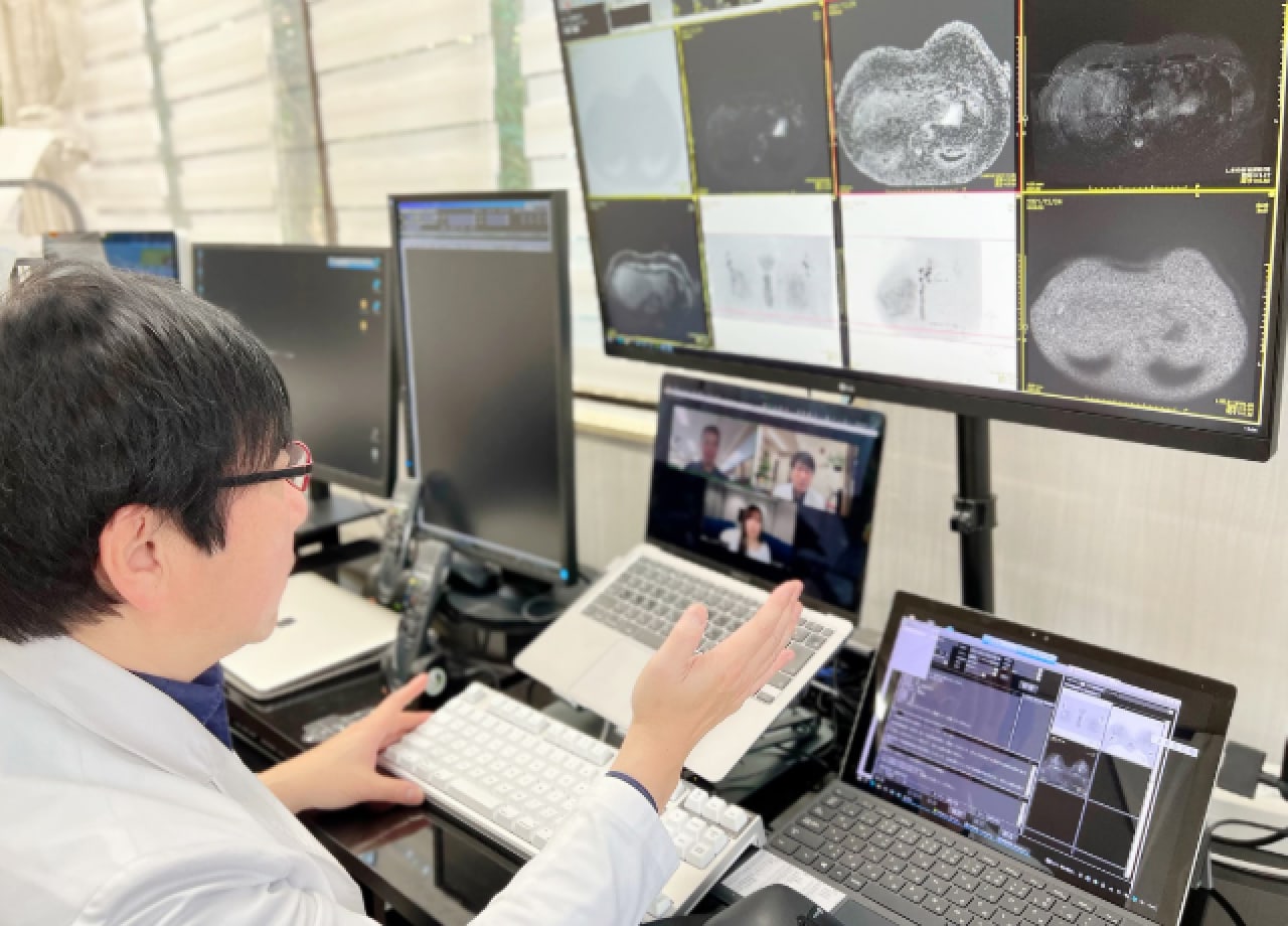
紹介先の乳腺外科への説明サービス
私共は、「貴院に乳腺外科がいなくても大丈夫です」と述べるだけではありません。サービスを導入していただいた病院様に対しては、近隣の乳腺外科(紹介先)をご紹介いただければ、その乳腺外科の先生にzoomでご説明するサービスも行っています。
06
受診者の満足度が高く、
家族や友達への
紹介につながります

極めて高い顧客推奨度(NPS)
受診者の顧客推奨度(NPS)は、驚異の+36.2(n=5815)。NPSは、それがプラスになればリピーターが増えると言われる厳しい指標ですが、この値は日本のすべての業種のトップレベルの値です。
もともとの「痛くない・見られない・スピーディ」という価値観だけでなく、アンケートを取り、その結果をフィードバックすること、また画質を常に安定させるように、サービス開始後も常に撮像現場にフィードバックすることによりこの値が支えられています。
07
導入費用が安く、
具体的なシミュレーション
を提示します

初期投資を短期間で回収可能
導入費用が安いため、モデル例では3ヶ月程度で初期費用を回収できます。黒字がでるための受診者数は週に1.5人足らずですので、単月黒字になるための損益分岐点は非常に低く、安心して導入いただけます。
これは当社が、いたずらに利益を追求する会社ではなく、乳がん検診時に受ける女性の痛みをなくしたい、それを普及させたいという理念に基づくもので、47都道府県すべてへの導入を目指しています。
DWIBS法の開発者の紹介

高原 太郎 医師
1961年東京都生まれ。秋田大学医学部卒業。聖マリアンナ医科大学放射線科勤務、東海大学医学部基盤診療学系画像診断学講師、オランダ・ユトレヒト大学病院放射線科客員准教授などを経て、2010年から東海大学工学部医用生体工学科教授
第32回日本乳癌検診学会
(45分)
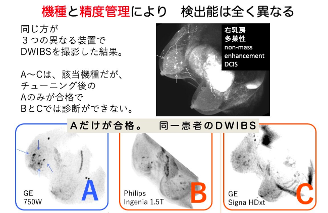
使用機種を絞り、
チューニングして実現。

従来MRI装置は、磁場強度(テスラ)だけが性能を示すと考えられてきました。
しかし実験してみると、機種や調整の有無で全く画質が異なり、診断に支障がでることがわかりました。
そこで、画質の性能検証が終わった2つのベンダーのMR装置のみを対象とし、画質調整後にサービス開始をしております。
その他の会社製については画質のチェックが終わり次第、順次対応をさせて頂く予定ですのでお待ち下さい。
※ キャノン最新型(Orian / Centurian)導入の方は、可能性がありますのでお問い合わせください(テストが必要です)。
これまでの診断成績
2019年3月までの1091例の集計において、がん発見率14.7%*、陽性適中率15.1%、要精査率9.7%でした。(MammographyおよびUSの参照なしの、非造影MRI単独読影での数値です)。
受診者背景が異なるため一概に比較できませんが、これはEVA trialで示されているCancer Yield (1000人あたりの発見数) で、Mammography+USの併用(7.7) の倍近い数値であり、かつ造影MRI (14.9)に近い値を示しています。
この高い診断成績は、「MRI自由自在」(メジカルビュー社)で示したような、Short T1, Short T2 changeに関する知識や、STIR/CHESSのlesion/backgroundコントラストの違い、TRの保持によるlong T1 lesion enhancementなどの知識を用いて画質を保持し、また診断することにより支えられています。
*patient baseなので実際のがん発見数はこれより多くなります。日本乳癌検診学会で発表予定です。
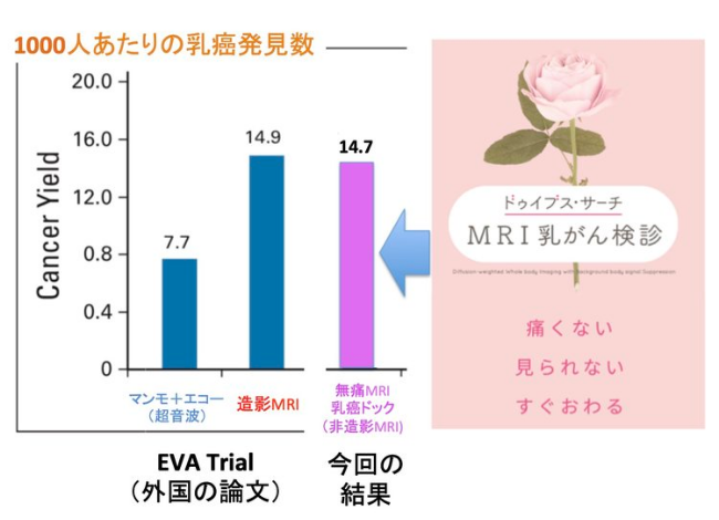
※ EVA Trialと 今回の無痛MRI乳がん検診の受診者背景は異なりますので直接比較はできないことにご注意ください。ただし今回のデータは1000例を超えた規模による集計です。
参考文献
・DWIBS法(第一報)
Takahara T, Imai Y, Yamashita T, Yasuda S, Nasu S, Van Cauteren M. Diffusion weighted whole body imaging with background body signal suppression (DWIBS): technical improvement using free breathing, STIR and high resolution 3D display. Radiat Med. 2004 Jul-Aug;22(4):275-82.
[983回被引用 (Google Scholar 2018.5)]
・DWIBS法(REVIEW, EUR RADIOL)
Kwee TC, Takahara T, Ochiai R, Nievelstein RA, Luijten PR. Diffusion-weighted whole-body imaging with background body signal suppression (DWIBS): features and potential applications in oncology. Eur Radiol. 2008 Sep;18(9):1937-52.
[413回被引用 (Google Scholar 2018.5)]
・DWIとDWIBS法の比較 (MAGN RESON IMAGING)
Mesmann C, Sigovan M, Berner LP, Abergel A, Tronc F, Berthezène Y, Douek P, Boussel L. Evaluation of image quality of DWIBS versus DWI sequences in thoracic MRI at 3T. Magn Reson Imaging. 2014 Dec;32(10):1237-41.
RESULTS: Quality of fat suppression was significantly higher for DWIBS than for DWI both for free-breathing (score 3.48±0.65 vs. 1.76±0.96, p<0.0001) and respiratory-gated scans (3.17±0.77 vs. 1.72±0.73, p=0.0001). Similarly, artifacts were reduced with DWIBS (3.16±0.47 vs. 1.76±0.59, p<0.0001; 3.0±0.73 vs. 2.04±0.53, p=0.0001). Quantitative analysis showed higher STB with DWIBS (3.26±1.83 vs. 0.98±0.44, p<0.0001; 3.56±, 2.09 vs. 0.92±0.59, p<0.0001). Gating did not improve image quality and STB on DWIBS (p>0.05).
・CANCER BIOMARKERとしてのDWIBS(NEOPLASIA)
Padhani AR, Liu G, Koh DM, Chenevert TL, Thoeny HC, Takahara T, Dzik-Jurasz A, Ross BD, Van Cauteren M, Collins D, Hammoud DA, Rustin GJ, Taouli B, Choyke PL.Diffusion-weighted magnetic resonance imaging as a cancer biomarker: consensus and recommendations. Neoplasia. 2009 Feb;11(2):102-25.
[1456回被引用 (Google Scholar 2018.5)]
DWIのうち、DWIBS法が適切な方法として推奨されている。
・DWIBS法による全身MR-NEUROGRAPHY (NEJM)
Yamashita T, Kwee TC, Takahara T. Whole-body magnetic resonance neurography. N Engl J Med. 2009 Jul 30;361(5):538-9.
DWIBS法による均一な背景信号抑制により、世界で初めて全身の末梢神経走行を描出し得た。
・DWIBS法と造影MRIによる乳がん検出能の比較(ガイドライン策定以降)
- 2015年(MAGN RESON IMAGING)
Telegrafo M, Rella L, Stabile Ianora AA, Angelelli G, Moschetta M. Unenhanced breast MRI (STIR, T2-weighted TSE, DWIBS): An accurate and alternative strategy for detecting and differentiating breast lesions. Magn Reson Imaging. 2015 Oct;33(8):951-5.
RESULTS: Unenhabced (UE)-MRI sequences obtained sensitivity, specificity, diagnostic accuracy, PPV and NPV values of 94%, 79%, 86%, 79% and 94%, respectively. CE-MRI sequences obtained sensitivity, specificity, diagnostic accuracy, PPV and NPV values of 98%, 83%, 90%, 84% and 98%, respectively. No statistically significant difference between UE-MRI and CE-MRI was found.
- 2016年(RADIOLOGY)
Bickelhaupt S, Laun FB, Tesdorff J, Lederer W, Daniel H, Stieber A, Delorme S, Schlemmer HP. Fast and Noninvasive Characterization of Suspicious Lesions Detected at Breast Cancer X-Ray Screening: Capability of Diffusion-weighted MR Imaging with MIPs. Radiology. 2016 Mar;278(3):689-97.
RESULTS: Twenty-four of 50 participants had a breast carcinoma. With AP1 (DWIBS), the sensitivity was 0.92 (95% confidence interval [CI]: 0.73, 0.98), the specificity was 0.94 (95% CI: 0.77, 0.99), the negative predictive value (NPV) was 0.92 (95% CI: 0.75, 0.99), and the positive predictive value (PPV) was 0.93 (95% CI: 0.75, 0.99). The mean reading time was 29.7 seconds (range, 4.9-110.0 seconds) and was less than 3 seconds (range, 1.2-7.6 seconds) in the absence of suspicious findings on the DWIBS MIPs. With the AP2 protocol, the sensitivity was 0.85 (95% CI: 0.78, 0.95), the specificity was 0.90 (95% CI: 0.72, 0.97), the NPV was 0.87 (95% CI: 0.69, 0.95), the PPV was 0.89 (95% CI: 0.69, 0.97), and the mean reading time was 29.6 seconds (range, 6.0-100.0 seconds).
- 2018年(JPN J RADIOLOGY)
Yamada T, Kanemaki Y, Okamoto S, Nakajima Y. Comparison of detectability of breast cancer by abbreviated breast MRI based on diffusion-weighted images and postcontrast MRI. Jpn J Radiol. 2018 May;36(5):331-339.
RESULTS: The study included 87 patients with 89 breast cancer lesions ≤ 2 cm in diameter. The sensitivity/specificity for AP1 (The abbreviated protocols based on DWI) and AP2 (postcontrast MRI) for reader 1 was 89.9/97.6% and 95.5/90.6%, respectively, and those for reader 2 was 95.5/94.1% and 98.9/94.1%, respectively. The AUCs for AP1 and AP2 for reader 1 were 0.9629 and 0.9640 (p = 0.95), respectively, and those for reader 2 were 0.9755 and 0.9843 (p = 0.46), respectively.
国内施設からの最新論文。Kuhl教授の唱える短縮MRI(Abbreviated MRI)の手法で読影比較をした結果でも、DWI(本研究ではDWIBSを用いている)と造影MRIで差がないことが示された。
・GD造影剤の沈着
*4 Kanda T, Nakai Y, Oba H, Toyoda K, Kitajima K, Furui S. Gadolinium deposition in the brain. Magn Reson Imaging. 2016 Dec;34(10):1346-1350. Review.
Gadolinium is highly toxic. Gadolinium-based contrast agents (GBCAs) consist of gadolinium ions and a chelating agent that binds the gadolinium ion tightly in order not to manifest its toxicity. Knowledge regarding gadolinium deposition in patients with normal renal function has advanced dramatically. Since 2014, increasing attention has been given to residual gadolinium known to accumulate in the tissues of patients with normal renal function. High signal intensity on T1-weighted images (T1WI) in the dentate nucleus, globus pallidus, and pulvinar region of the thalamus correlate roughly with the number of previous GBCA administrations. Pathological analyses have revealed that residual gadolinium is deposited not only in these brain regions, but also in extracranial tissues such as liver, skin and bone. The risks attendant with these deposits are unknown. Common sense dictates that gadolinium deposition be kept as low as possible, and that gadolinium contrast agents be used only when absolutely necessary, with preferential use of macrocyclic chelates, which seem to be deposited at lower concentrations.
2014年、MRI用 造影剤(ガドニウム、Gd)は脳に沈着すること、使用するほど沈着の度合いが高くなること(dose dependent:用量依存性)が報告された。その後の研究で2種類の造影剤(リニア型、マクロ環型)のうち、リニア型は10倍の沈着を招くことが判明。2017年8月に欧州で発売中止、同11月末に厚生労働省より「脳への残存が報告されていることを踏まえ、ガドリニウム造影剤を用いた検査の必要性を慎重に判断する。線状型は環状型より残存しやすいことが報告されていることを踏まえ、環状型 の使用が適切でない場合に投与する」と通知される [リンク]。
よくある質問
どこで受けられますか?
乳房インプラントが入っていますが受けられますか?
どのぐらいの時間が必要ですか? なにか準備は必要ですか?
どのぐらいがんが見つかるのですか?
生理や妊娠・授乳との関係はありますか?
被ばくはないでしょうか?
MRI乳がん検診はどうやってがんを見つけるのですか?
MRI乳がん検診はどのくらい(回数・頻度)受ければいいですか?
閉所恐怖なのですが大丈夫でしょうか?
MRI乳がん検診を受けることができない場合はありますか?
MRIはどんな音がしますか?
- 全国の病院を探す |
- 無痛MRI乳がん検診の流れ |
- 痛くない理由 |
- 見られない理由 |
- 被ばくゼロの理由 |
- がん発見率の高い理由
- 乳房手術後も検査可能な理由 |
- 日本人に適している理由 |
- 検査結果報告書 |
- 乳がんってどんな病気? |
- 乳がん検診の種類
- マンガで見る無痛MRI乳がん検診 |
- 動画で見る無痛MRI乳がん検診 |
- 体験者の声 |
- 開発者の想い |
- よくある質問
- 医療関係・メディア関係のみなさまへ |
- 運営機関


